

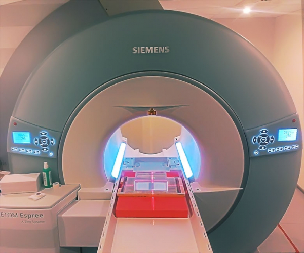
A screwdriver whizzed past my ear. With a loud clang, it stopped on the body of the cryostat. Cursing to myself, I decided to take a break. Unscrewing bolts in a magnetic field of 1.5 tesla using a steel tool is not the best idea. The field, like an invisible opponent, constantly tries to yank the tool from my hands, align it along its lines of force, and pull it as close as possible to the electrons racing around the superconducting loop. However, if you really need to tackle the corroded connections from years past, there's not much choice. I sat down at the computer and habitually scrolled through the news feed. "Russian scientists have improved MRI by 2 times!" read the suspicious headline.
About a year ago, we and delved into understanding how it works. I highly recommend refreshing your memory with that material before reading this article.
Due to various reasons, including historical ones, in Russia today there is production of such complex equipment as high-field magnetic resonance tomographs. Nevertheless, if you live in a relatively large city, you will easily find clinics offering such services. Moreover, the MRI scanner fleet is often represented by used equipment that was once imported from the USA and Europe, and if you happen to visit a clinic with an MRI, don't be deceived by the device's shiny appearance — it might very well be entering its second decade. Consequently, such equipment can break down, and for a long time, I was one of those people who brought broken tomographs back to life so that patients could continue to receive diagnoses and owners could keep making profits.
One fine day, during a break between dangerous activities with massive magnetic fields, I stumbled upon an interesting note in the news feed: "Russian scientists, together with their Dutch colleagues, using metamaterials." It is worth mentioning that the very fact that research is being conducted in Russia on equipment whose production has not yet been mastered seemed to me quite controversial. I thought it was just another instance of grant misappropriation, diluted with incomprehensible scientific jargon like the already tiresome concept of "nanotechnology." My search for information on the work of domestic scientists with MRI and metamaterials led me to an article describing a simple experiment that I could easily replicate, as I always have an MRI machine on hand.
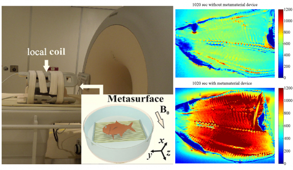
Image from , dedicated to enhancing MRI signals using a so-called "metamaterial." In a typical clinical 1.5-Tesla machine, instead of a patient, a metamaterial is loaded in the form of a basin of water, inside of which parallel wires of a specific length are located. An object of study—a fish (non-living)—is placed on the wires. The images on the right are MRI scans of the fish, overlaid with a color map indicating the intensity of the signal from hydrogen nuclei. It is clear that when the fish is lying on the wires, the signal is much better than without them. The scanning time in both cases is the same, which proves the increase in scanning efficiency. The article also conveniently provided
a formula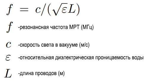
to calculate the length of the wires depending on the operating frequency of the tomograph, which I utilized. I crafted my metamaterial from a cuvette and a set of copper wires, securing them with plastic fasteners printed on a 3D printer:
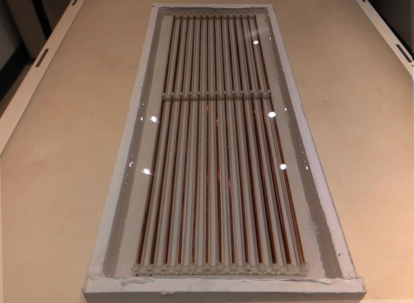
My first metamaterial. Immediately after fabrication, it was inserted into a 1-Tesla tomograph. An orange served as the scanning object.
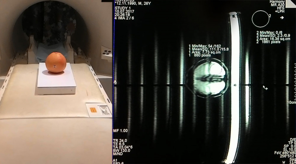
However, instead of the promised signal enhancement, I received a pile of artifacts that completely spoiled the image! My frustration knew no bounds! After finishing the specimen, I wrote a letter to the authors of the article, the essence of which can be summed up with the question "What the... ?".
The authors responded to me quite quickly. They were rather impressed that someone was trying to replicate their experiments. At first, they spent a long time trying to explain how metamaterials work, using terms like "Fabry-Perot resonances," "normal modes," and various radio frequency fields. Then, apparently realizing that I had no idea what they were talking about, they decided to invite me to their place so that I could see their developments in person and confirm that it actually works. I packed my favorite soldering iron in my backpack and headed to St. Petersburg, to the National Research University of Information Technologies, Mechanics and Optics (it turned out that they teach not only programmers there).

Upon arrival, I was warmly welcomed, and suddenly, they offered me a job, as they were impressed by my cuvette with wires and needed someone to create new ones. In return, they promised to explain everything that interested me in detail and to take me through a course on radio physics and MRI, which, by a happy coincidence, was starting that very year. My thirst for knowledge won out, and over the next year, I studied, worked on projects, and gradually learned more and more about the history of magnetic resonance as well as the state of modern science in this field, which I will share here.
At the core of the proposed method for improving MRI, and explored in the mentioned scientific articles, are so-called "metamaterials." Metamaterials, like many other discoveries, owe their existence to unexpected solutions derived from theoretical research. The Soviet scientist, Viktor Veselago, in 1967, while working on a theoretical model, hypothesized the existence of materials with a negative refractive index. As you may have gathered, this pertains to optics, and this coefficient roughly indicates how much the direction of light will change as it passes through the boundary between different mediums, such as air and water. You can easily confirm that this indeed happens yourself:
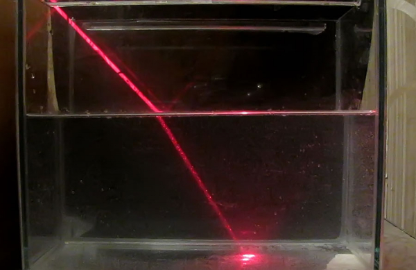
A simple experiment with a laser pointer and an aquarium demonstrating light refraction.
An interesting fact that can be derived from such an experiment is that a beam cannot refract in the same direction from which it fell on the boundary, no matter how hard the experimenter tries. Such experiments have been conducted with all substances found in nature, yet the beam stubbornly refracted only in one direction. Mathematically, this means that the index of refraction, along with its components, the dielectric and magnetic permeability, are positive, and no other observation has been made. At least, not until V. Veselago decided to study this question and showed that theoretically there is no reason why the index of refraction couldn't be negative.
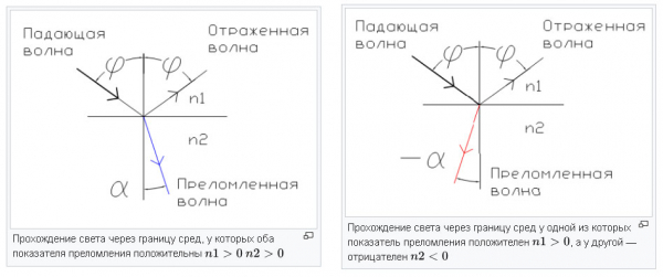
An image from Wikipedia showing the difference between media with positive and negative refractive indices. As we can see, light behaves completely unnaturally compared to our everyday experience.
For a long time, V. Veselago tried to find evidence for the existence of materials with a negative index of refraction, but his search was unsuccessful, and his work was unjustly forgotten. Only at the beginning of the next century were composite structures artificially created that realized the described properties, but not in the optical range, rather in a lower microwave frequency range. This became a turning point, as the very possibility of such materials opened new perspectives. For example — the creation of , capable of enlarging objects even smaller than the wavelength of light. Or — absolute cloaking coatings, the dream of all military applications. Significant revisions were made to the theory to account for new data. The key to success turned out to be the use of ordered structures made of resonant elements — meta-atoms, whose size is much smaller than the wavelength of radiation with which they interact. An ordered structure of meta-atoms is an artificial composite called metamaterial.
The practical implementation of metamaterials is technologically complex even today, as the size of the resonant particles must be comparable to the wavelength of electromagnetic radiation. For the optical range (where the wavelength is in nanometers), such technologies are at the forefront of progress. Therefore, it's not surprising that the first representatives of the metamaterial concept were created for relatively longer electromagnetic waves in the radio range (which have a more familiar length from mm to m). The main feature and at the same time drawback of any metamaterial is a result of the resonant nature of its constituent elements. A metamaterial can exhibit its miraculous properties only at certain frequencies.
Limited frequencies.So, for example, the next time you see something like a super sound blocker based on metamaterials, ask what frequency range it actually blocks.
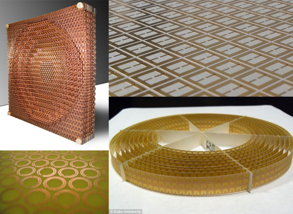
Typical examples of metamaterials that allow interaction with electromagnetic waves. Structures made of conductors are nothing more than small resonators, LC circuits formed by the spatial arrangement of conductors.
It has been a little while since the concept of metamaterials and their first implementations appeared, and people figured out how to use them in MRI. The main drawback of metamaterials — the narrow operational range — is not a problem for MRI, where all processes occur almost at the same frequency of magnetic resonance of nuclei, which lies in the radio range. Here you can create meta-atoms with your own hands and immediately see what they look like in images. Among the first features that researchers implemented in MRI using metamaterials were superlenses and endoscopes.
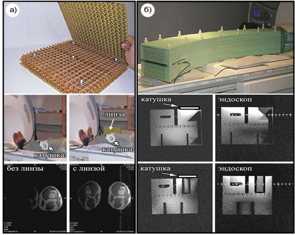
On the left side under section a), a superlens is depicted, consisting of a three-dimensional lattice of resonators on printed circuit boards. Each resonator is an open metallic ring with a soldered capacitor, forming an LC circuit tuned to the MRI frequency. Below is an example of how this metamaterial structure is placed between the patient's legs during the tomography procedure, along with the resulting images. If you didn't hesitate to read my previous article on MRI, you already know that capturing an image of any part of a patient's body requires collecting weak, rapidly decaying signals from nuclei using a closely positioned antenna – a coil.
The metamaterial superlens allows for an expanded coverage area of the standard coil. For example, it can visualize both legs of the patient simultaneously instead of just one. The downside is that the position of the superlens must be carefully adjusted to achieve the best effect, and the superlens itself is quite expensive to manufacture. If you still don't understand why this lens is referred to as 'super-', take a look at its size in the photo, and then realize that it works with a wavelength of about five meters!
Under section b), the structure of the endoscope is demonstrated. Essentially, the endoscope for MRI is an array of parallel wires, acting as a waveguide. It allows for spatial separation between the region from which the coil receives signals from nuclei and the coil itself, to the extent that the receiving antenna can be located entirely outside the cryostat of the tomograph, far from the permanent magnetic field. The images shown in section b) below were obtained for a specially liquid-filled vessel – a phantom. The difference between them is that the images labeled 'endoscope' were captured when the coil was placed at a considerable distance from the phantom, where without the endoscope, signals from the nuclei would have been impossible to detect.
When discussing one of the most promising applications of metamaterials in MRI, and one that is closest to practical implementation (which I ultimately got involved in) – it is the creation of wireless coils. It's worth explaining that this is not about Bluetooth or any other wireless data transmission technology. 'Wireless' in this case refers to the presence of inductive or capacitive coupling between two resonant structures – a transmitting and receiving antenna, as well as the metamaterial. Conceptually, it looks like this:
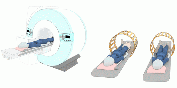
On the left, the typical MRI procedure is shown: the patient lies inside the cryostat within a uniform static magnetic field. A large antenna, called a 'birdcage', is mounted in the tunnel of the tomograph. This configuration allows the rotation of the radiofrequency magnetic field vector at the precession frequency of hydrogen nuclei (for clinical machines, this is usually between 40 and 120 MHz, depending on the strength of the static magnetic field from 1T to 3T respectively), causing them to absorb energy and subsequently emit a response. The response signal from the nuclei is very weak, and by the time it reaches the conductors of the large antenna, it inevitably attenuates. For this reason, MRI uses nearby local coils to receive signals. The center image, for example, shows a typical situation in knee scanning. With the use of metamaterials, a resonator can be created that will be inductively coupled with the birdcage. It is enough to place such a device near the required area of the patient's body, and the signal from there will be received just as well as with a local coil! If the concept is successfully implemented, patients will no longer have to deal with wires, making the MRI diagnostic procedure more comfortable.
This is exactly what I tried to create at first, pouring water over wires and attempting to scan an orange. The wires submerged in water from the very first image in this article are nothing other than meta-atoms, each of which acts as a half-wave dipole – one of the most well-known antenna designs familiar to every radio enthusiast.
They immerse them in water not to prevent them from catching fire during MRI (although that's part of it too), but to reduce their resonance length thanks to the high dielectric constant of water by an amount equal to the square root of the dielectric constant of water.
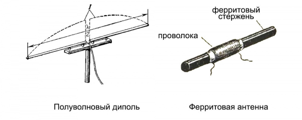
This technique has long been used in radio receivers by winding wire around a piece of ferrite — the so-called ferrite antenna. However, ferrite has a high magnetic permeability rather than a dielectric, which still works and allows for the reduction of the antenna's resonance sizes. Unfortunately, you can't use ferrite in MRI because it is magnetic. Water is a cheap and accessible alternative.
It's clear that to calculate all these things, complex mathematical models need to be constructed, taking into account the interconnections between resonance elements, environmental parameters, and radiation sources... or you can take advantage of the advancements in software for numerical electromagnetic modeling, which even a school student can easily handle (bright examples include CST, HFSS). The software allows you to create 3D models of resonators, antennas, electrical circuits, adding in people – in fact, anything you like; it just depends on your imagination and available computational power. The constructed models are divided into grids, at the nodes of which the known Maxwell's equations are solved.
For example, here is a simulation of the radio frequency magnetic field inside the previously mentioned birdcage-type antenna:
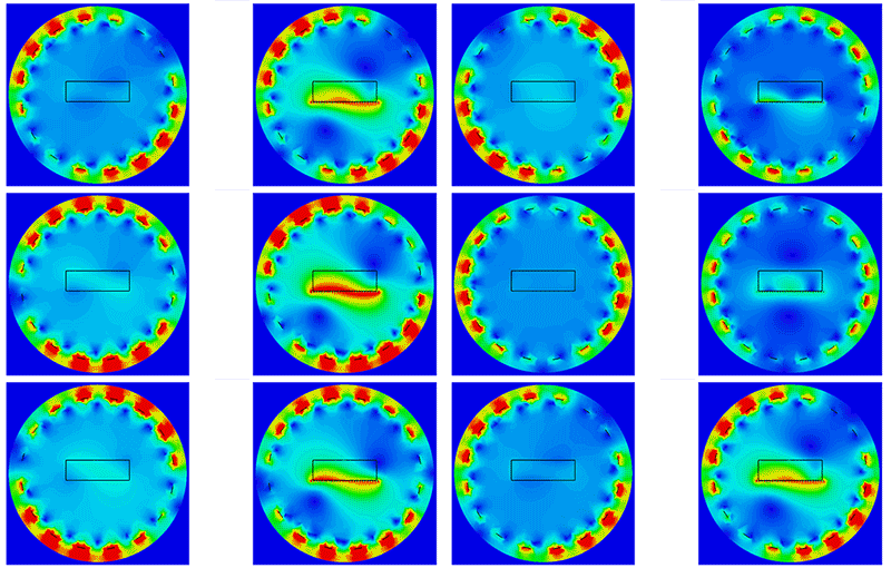
It becomes quite evident how the field rotates. On the left is the situation when the antenna contains a box of water, while on the right is the same box on a wire resonator of resonant length. You can see how the magnetic field is significantly enhanced by the wires. After mastering CST and optimizing my design there, I created a metamaterial that truly allowed for signal enhancement in a standard 1.5T clinical MRI tomograph. It still represented a box (though a more aesthetically pleasing one made of plexiglass), filled with water and a mass of wires. This time, the structure was optimized in terms of resonance conditions, namely: selecting wire lengths, their placements, and the amount of water. Here’s what I got with the tomato:
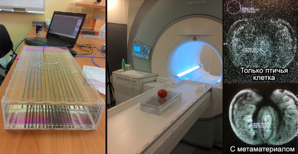
The first scan of the tomato was performed on a large antenna. As a result, there was only noise with barely discernible outlines. The second time, I placed the fruit on a freshly baked resonant structure. I didn’t create colorful maps or anything similar, as the effect was evident. Thus, from my experience, albeit spending a lot of time, I proved that the concept works.
I understand what you're thinking — oranges, tomatoes — that’s not it, where are the human trials?
They were indeed :
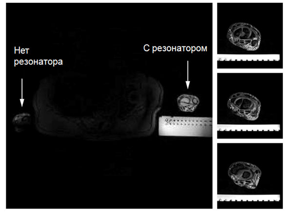
The hand of a volunteer undergoing an MRI lies on the same box. The water in the box, because it contains hydrogen, is also clearly visible. Signal enhancement occurs in the wrist area lying on the resonator, while other body parts are poorly visible. It’s clear that such an effect, or perhaps even better, can be achieved using standard clinical coils. But the fact that such things can be done simply by spatially combining water and wires in the right way is mind-blowing. Even more astonishing is that knowledge about this can be gained by studying seemingly unrelated phenomena, such as the refraction of light.
For those who are still not tiredCurrently, the design of the box with water has already been improved. Now it’s just a flat printed circuit board, which allows localizing the magnetic field of the external large antenna around it. Moreover, its working area is larger than that of the previous design:
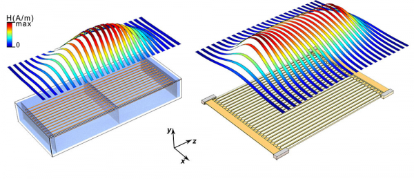
Colored ribbons show the intensity of the magnetic field above the structure when excited by an external source of electromagnetic waves. The flat structure represents a typical transmission line known in radio engineering, but it can also be regarded as a metamaterial for MRI. This "wireless coil" can already compete with standard coils in uniformity of the generated field at a certain depth in the scanning object:
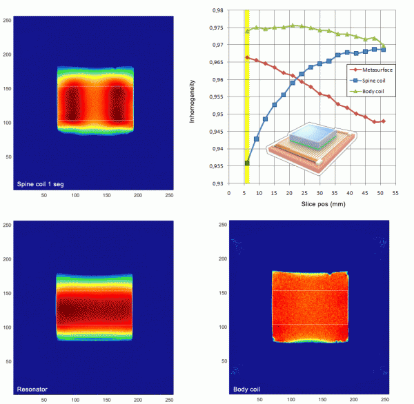
The animation shows a layered color map of the signal inside a water box in an MRI. The color indicates the intensity of signals from hydrogen nuclei. In the upper left corner, a segment of a standard coil is used as the receiver for scanning the back. The lower left corner shows the box resting on a resonator in the shape of a circuit board. In the bottom right, a large antenna built into the tomograph tunnel receives the signal. I compared the uniformity of the signal in the area outlined in a rectangle. At a certain height, the metamaterial performs better than the coil in terms of signal uniformity. While this may not be crucial for clinical tasks, when it comes to scientific MRI setups where rats are scanned, it can help achieve signal gains and reduce the required power of the excitation radio impulses.
The claim of "twice as good" at the start of the article is, of course, another product of the journalists' unreciprocated affection for scientists. However, it would also be wrong to say that these studies are empty, as evidenced by the interest in this topic among research groups worldwide. Surprisingly, work is also being done here in Russia, although from my purely personal experience, this is more of a rare exception. There are still many unresolved problems associated with the application of metamaterials in MRI. In addition to localizing magnetic fields for good imaging, one should not forget about the electric fields that lead to tissue heating, as well as the absorption of energy from the radiofrequency field by patients undergoing examination. There should be special control over these factors in clinical use, which becomes significantly more complicated with the use of field-localizing resonators. Currently, metamaterials for MRI remain within the framework of scientific research, but the results obtained are already quite interesting, and it is possible that in the future, the MRI procedure will change for the better, becoming faster and safer.
Source: habr.com
