

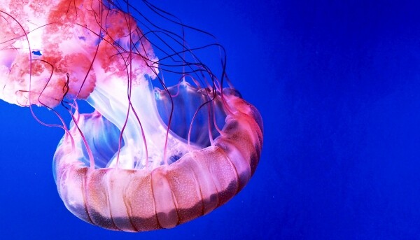
What do Wolverine, Deadpool, and jellyfish have in common? They all possess a remarkable ability — regeneration. Of course, in comics and movies, this ability, which is common among a very limited number of real living organisms, is slightly (and sometimes significantly) exaggerated, but it remains quite real. And what is real can be explained, which is what scientists from Tohoku University (Japan) have set out to do in their new study. What cellular processes in the jellyfish's body are linked to regeneration, how does this process occur, and what other super abilities do these gelatinous creatures possess? This will be revealed by the research group's report. Let's go.
The foundation of the research
First of all, the scientists explain why they chose to focus on jellyfish. The fact is that most biological research is conducted using so-called model organisms: mice, fruit flies, worms, fish, etc. But our planet is home to millions of species, each possessing their own unique abilities. Therefore, it is impossible to fully evaluate the process of cellular regeneration by studying just one species and assuming that the mechanism studied will be universal for all life on Earth.
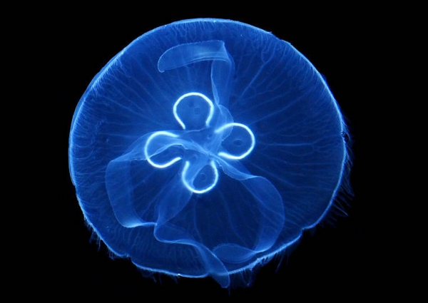
As for jellyfish, these creatures speak of their uniqueness simply through their appearance, which cannot help but attract scientists' attention. Therefore, before delving into the dissection of the study itself, I got acquainted with its main character.
The word 'jellyfish', which we use to refer to the creature as such, actually only denotes a stage in the life cycle of cnidarians from the subphylum Medusozoa. Cnidarians got their unusual name due to the presence of stinging cells (cnidocytes) in their bodies, which are used for hunting and self-defense. Simply put, when you get stung by a jellyfish, you can thank these cells for the pain and suffering.
Cnidocytes contain nematocysts — intracellular organelles responsible for the 'stinging' effect. There are several types of cnidocytes distinguished by their appearance and, accordingly, their method of use, among which the following can be highlighted:
- Penetrants are threads with pointed ends that penetrate the body of the victim or aggressor like spears, injecting neurotoxin;
- Glutinants are sticky, long threads that envelop the victim (not the most pleasant hugs);
- Volvents are short threads in which the victim can easily become entangled.
This unconventional weaponry is explained by the fact that jellyfish, while graceful, are not particularly quick creatures. The neurotoxin that enters the prey’s body instantly paralyzes it, giving the jellyfish plenty of time for its lunch break.
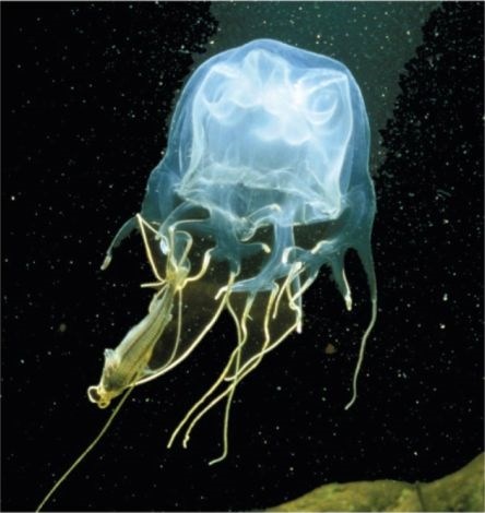
A jellyfish after a successful hunt.
In addition to their unusual hunting and defense methods, jellyfish also have a very peculiar reproductive process. Males produce sperm, while females produce eggs, which, after fusion, form planulas (larvae) that settle to the bottom. Over time, the larvae develop into polyps, from which young jellyfish literally break off when they reach maturity (in fact, this is budding). Thus, there are several stages in the life cycle, one of which is the jellyfish or medusoid generation.
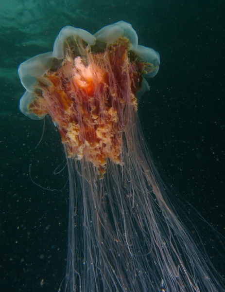
The hairy cyanide, also known as the 'lion’s mane.'
If the hairy cyanide were asked how to increase hunting efficiency, it would respond — more tentacles. There are about 60 of them (clusters of 15 tentacles at each corner of the umbrella). Additionally, this jellyfish species is considered the largest, as its umbrella diameter can reach 2 meters, and its tentacles can extend up to 20 meters during hunting. Fortunately, this species is not particularly 'venomous,' and therefore not lethal to humans.
The box jellyfish, in turn, would add quality to the quantity. This type of jellyfish also has 15 tentacles (3 m long) on each of the four corners of its bell, but its venom is many times stronger than that of its larger relative. It is believed that the neurotoxin in the box jellyfish’s body is enough to kill 60 people in 3 minutes. This terror of the seas resides in the coastal waters of northern Australia and New Zealand. According to data from 1884 to 1996, 63 people died in Australia, but these figures may be inaccurate, and the number of fatal encounters between humans and box jellyfish could be much higher. However, data from 1991 to 2004 indicate that among 225 cases, only 8% of the victims were hospitalized, including one fatal outcome (a three-year-old child).

Box Jellyfish
Now let’s return to the study we are discussing today.
From a cellular perspective, the most important process in the life of any organism is cellular proliferation — the process of tissue growth through cell division. During the growth of an organism, this process regulates body size increase. When the organism is fully formed, proliferating cells regulate physiological cell turnover and replace damaged ones with new cells.
Cnidarians, being a related group of bilateral and early branches in the evolution of multicellular organisms, have been used to study evolutionary processes for many years. Therefore, cnidarians are no exception when it comes to proliferation. For example, during the embryonic development of the sea anemone Nematostella vectensis cell proliferation is coordinated with epithelial organization and contributes to tentacle development.
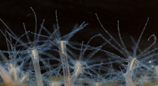
Nematostella vectensis
Moreover, as we already know, cnidarians are known for their regenerative abilities. Among researchers, hydra polyps (a genus of freshwater sessile cnidarians from the class Hydroida) have been popular for hundreds of years. Proliferation activated by dying cells initiates the regeneration process of the hydra's basal head. The very name of this creature hints at the mythical being known for its regeneration — the Lernaean Hydra, which Hercules was able to defeat.
While regenerative capabilities have been linked to proliferation, it remains unclear how exactly this cellular process occurs under normal conditions at different stages of organism development.
Jellyfish, which have a complex life cycle consisting of two reproductive stages (asexual and sexual), serve as an excellent model for studying proliferation.
In this study, the main researched subject was the jellyfish species Cladonema pacificum. This species inhabits the shores of Japan. Initially, this jellyfish has 9 primary tentacles, which begin to branch and increase in size (just like the rest of the body) during development to adulthood. This feature allows for a detailed study of all the mechanisms involved in this process.
In addition to Cladonema pacificum the study also examined other jellyfish species: Cytaeis uchidae and Rathkea octopunctata.
Research Results
To understand the spatial pattern of cell proliferation in the Cladonema medusa, scientists applied 5-ethynyl-2'-deoxyuridine (EdU) staining, which labels cells in the S-phase* or cells that have already passed through it.
S-phase* — the phase of the cell cycle in which DNA replication occurs.
Considering that Cladonema dramatically increases in size and demonstrates tentacle branching during development (1A—1C), the distribution of proliferating cells may change throughout maturation.
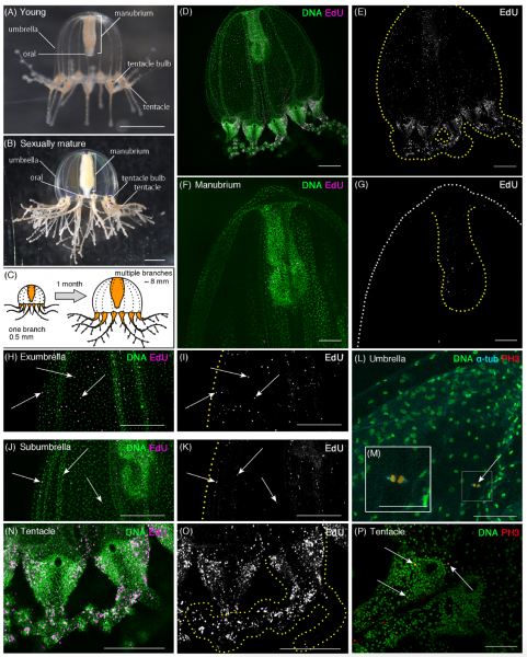
Image No. 1: characteristics of cell proliferation in young Cladonema.
Due to this feature, it was possible to study the mechanism of cell proliferation in both young (day 1) and mature (day 45) jellyfish.
In young jellyfish, EdU-positive cells were found in large numbers throughout the body, including the bell, manubrium (the supporting organ of the mouth in jellyfish), and tentacles, regardless of the exposure time to EdU (1D–1K and 1N–1O, EdU: 20 µM (micromolar) after 24 hours).
In the manubrium, very few EdU-positive cells were found (1F and 1G), while their distribution in the bell was very even, especially in the outer layer of the bell (exumbrella, 1H—1K). In the tentacles, EdU-positive cells were highly clustered (1N). The use of the mitotic marker (anti-PH3 antibodies) confirmed that EdU-positive cells are indeed proliferating cells. PH3-positive cells were found both in the bell and in the bulb of the tentacle (1L and 1P).
In tentacles, mitotic cells were primarily found in the ectoderm (1P), while proliferating cells in the umbrella were located in the surface layer (1M).
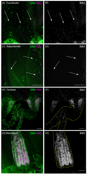
Image #2: features of cellular proliferation in mature Cladonema.
As with young individuals, numerous EdU-positive cells were found throughout the body of mature specimens. In the umbrella, EdU-positive cells were more frequently located in the surface layer than in the lower layer, similar to observations in younger individuals (2A—2D).
However, in the tentacles, the situation was somewhat different. EdU-positive cells accumulated at the base of the tentacle (bulb), where two clusters were identified on either side of the bulb (2E and 2F). In young individuals, similar accumulations were also observed (1N), meaning that the bulbs of the tentacles may be a primary area of proliferation throughout the medusoid stage. Interestingly, in the manubrium of adult specimens, the number of EdU-positive cells was significantly higher than in young ones (2G and 2H).
The intermediate conclusion is that cell proliferation may occur uniformly in the medusa's umbrella, whereas in the tentacles this process is highly localized. Consequently, it can be suggested that uniform cell proliferation may control body growth and tissue homeostasis, while clusters of proliferating cells near the bulbous base of the tentacles participate in the morphogenesis of the tentacles.
In terms of body development as such, proliferation plays a crucial role in body growth.
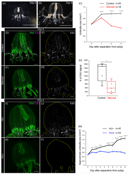
Image #3: the importance of proliferation in the process of medusa body growth.
To verify this in practice, researchers tracked the growth of medusa bodies starting from young individuals. The easiest way to determine the size of a medusa is by its dome, as it grows uniformly and directly proportionally to the entire body.
Under normal feeding conditions in the laboratory, the dome size increases sharply by 54.8% over the first 24 hours — from 0.62 ± 0.02 mm² to 0.96 ± 0.02 mm². In the following 5 days of observation, the size slowly and steadily increased to 0.98 ± 0.03 mm² (3A—3C).
Medusae from another group, which were deprived of food, did not grow but rather decreased in size (the red line on the graph 3C). Cellular analysis of starving medusae showed an extremely low number of EdU cells: 1240.6 ± 214.3 in medusae from the control group and 433.6 ± 133 in starving ones (3D—3H). This observation may be direct evidence that nutrition directly affects the proliferation process.
To test this hypothesis, scientists conducted a pharmacological analysis, during which they blocked cell cycle progression using hydroxyurea (CH4N2O2) — a cell cycle inhibitor that halts the G1 phase. As a result of this intervention, S-phase cells previously identified using EdU disappeared (3I—3L). Thus, the jellyfish exposed to CH4N2O2 did not demonstrate body growth, unlike the control group (3M).
The next stage of the study involved a detailed examination of the branching tentacles of jellyfish to confirm the assumption that local cell proliferation in the tentacles contributes to their morphogenesis.
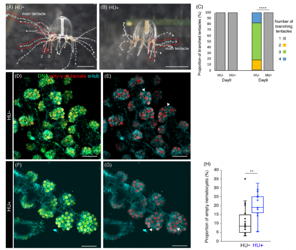
Figure 4: The effect of local proliferation on the growth and branching of jellyfish tentacles.
Young jellyfish tentacles have one branch, but their number increases over time. In laboratory conditions, branching increased threefold by the ninth day of observations (4A and 4C).
Again, using CH4N2O2, branching of the tentacles was not observed, and there was only one branch (4B and 4C). Interestingly, the removal of CH4N2O2 from the jellyfish restored the process of tentacle branching, indicating the reversibility of the pharmacological intervention. These observations clearly indicate the importance of proliferation for tentacle development.
Cnidarians would not be cnidarians without nematocysts (a cnidoct, i.e., stinging cells). In the jellyfish species Clytia hemisphaerica, stem cells in the tentacle bulbs supply nematocysts to the tips of the tentacles precisely through cell proliferation. Naturally, scientists decided to test this assertion.
To detect any connection between nematocysts and proliferation, a nuclear staining dye was used, which can mark poly-γ-glutamate synthesized in the wall of the nematocyst (DAPI, i.e., 4’,6-diamidino-2-phenylindole).
Staining of poly-γ-glutamate allowed for the estimation of nematocyst size, varying from 2 to 110 μm² (4D—4G). A certain number of empty nematocysts were also identified, meaning these nematocysts were depleted (4D—4G).
The proliferation activity in the tentacles of jellyfish was assessed by studying the vacuoles in the nematocysts after cell cycle blockage with CH4N2O2. The proportion of empty nematocysts in jellyfish after the drug intervention was higher than in the control group: 11.4% ± 2.0% in jellyfish from the control group and 19.7% ± 2.0% in jellyfish treated with CH4N2O2 (4D—4G and 4H). Therefore, even after depletion, nematocysts continue to be actively supplied with precursor cells for proliferation, confirming the influence of this process not only on tentacle development but also on nematogenesis within them.
The most interesting part of the study was the investigation of the regenerative abilities of jellyfish. Given the high concentration of proliferative cells in the tentacle bulb of mature jellyfish Cladonema, the researchers decided to study the regeneration specifically of the tentacles.
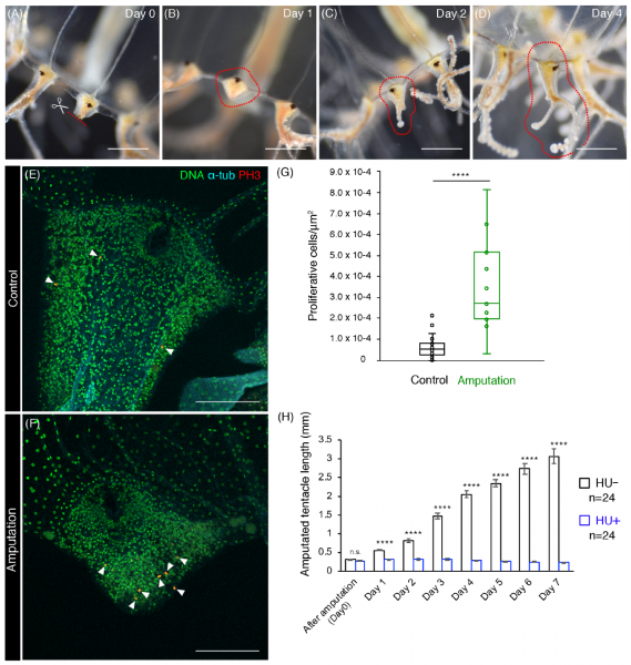
Figure 5: The effect of proliferation on tentacle regeneration.
After dissecting the tentacles at the base, a regeneration process was observed (5A–5D). During the first 24 hours, healing occurred at the cut site (5B). On the second day of observations, the tip began to lengthen and branches appeared (5C). By the fifth day, the tentacle was fully branched (5D), thus, the regeneration of the tentacle appears to follow normal morphogenesis after elongation.
To better study the initial stage of regeneration, the scientists analyzed the distribution of proliferating cells using PH3 staining to visualize mitotic cells.
While dividing cells were frequently observed near the amputated area, mitotic cells were dispersed in uncut control tentacle bulbs (5E and 5F).
). Quantitative assessment of PH3-positive cells present in the tentacle bulbs revealed a significant increase in PH3-positive cells in the tentacle bulbs of individuals with amputated limbs compared to the control group (5G). In conclusion, initial regenerative processes are accompanied by an active increase in cell proliferation in the tentacle bulbs.
The effect of proliferation on regeneration was assessed by blocking cells with CH4N2O2 after tentacle amputation. In the control group, the elongation of the tentacle after amputation occurred normally, as expected. However, in the group treated with CH4N2O2, elongation did not occur despite normal wound healing (5H). In other words, healing will occur in any case, but proliferation is essential for proper tentacle regeneration.
Finally, the researchers decided to study proliferation in other jellyfish species, specifically in Cytaeis and Rathkea.
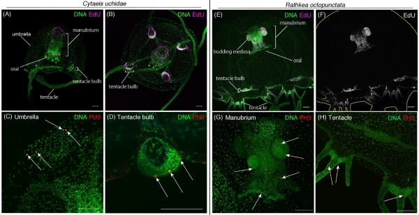
Image No. 6: comparison of proliferation in jellyfish species Cytaeis (on the left) and Rathkea (on the right).
The Cytaeis EdU-positive medusa cells were observed in the manubrium, tentacle bulbs, and the upper part of the bell (6A and 6B). The location of the identified PH3-positive cells in Cytaeis is very similar to Cladonema, but there are some differences (6C and 6D). In contrast, Rathkea EdU-positive and PH3-positive cells were found almost exclusively in the area of the manubrium and tentacle bulbs (6E—6H).
Interestingly, proliferating cells were often identified in the jellyfish buds Rathkea (6E—6G), reflecting the asexual reproduction type of this species.
Considering the information obtained, it can be assumed that cell proliferation occurs in the tentacle bulbs of more than just one jellyfish species, although there are differences due to variations in physiology and morphology.
For a more detailed understanding of the nuances of the research, I recommend checking out the .
Epilogue
One of my favorite literary characters is Hercule Poirot. The perceptive detective always paid special attention to small details that seemed unimportant to others. Scientists are much like detectives, gathering all the evidence they can find to answer every question of the investigation and identify the 'culprit.'
As obvious as it may sound, jellyfish cell regeneration is directly related to proliferation — an integral process in the development of cells, tissues, and consequently, the entire organism. A more meticulous study of this comprehensive process will allow us to better understand the molecular mechanisms underlying it, which in turn will expand not only our knowledge base but also have a direct impact on our lives.
Friday off-topic:
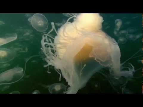
The march of jellyfish of the Aurelia species, disturbed by a predator with the unusual name "fried egg jellyfish" (Planet Earth, voiceover by David Attenborough).

Not a jellyfish, but this deep-sea creature (the pelican eel) is rarely captured on film (the researchers' reaction is simply heartwarming).
Thank you for your attention, stay curious, and have a great weekend, everyone! 🙂
Thank you for staying with us. Do you enjoy our articles? Want to see more interesting content? Support us by placing an order or recommending us to your friends, 30% discount for Habr users on a unique entry-level server designed by us for you: (options available with RAID1 and RAID10, up to 24 cores and up to 40GB DDR4).
Dell R730xd for half the price? Only with us in the Netherlands! Dell R420 — 2x E5-2430 2.2GHz 6C 128GB DDR3 2x960GB SSD 1Gbps 100TB — from $99! Read about how
Source: habr.com
