

Movement is life. This phrase can be interpreted as motivation to move forward, not to stand still and to achieve desired goals, as well as a statement of the fact that practically all living beings spend most of their lives in motion. In order for our movements and spatial displacements not to end with bumps on our foreheads and broken little toes, our brain uses stored "maps" of the environment that unconsciously surface at the moment of our movement. However, there is an opinion that the brain applies these maps not externally, so to speak, but by placing the individual on this map and gathering data from a first-person perspective. This theory was put to the test by scientists from Boston University, who conducted a series of practical experiments with laboratory rats. How does the brain actually orient itself in space, which cells are involved, and what role does this research play for the future of autonomous vehicles and robots? We will find out from the research group's report. Let's go.
The foundation of the research
So, the fact established many years ago is that the main part of the brain responsible for spatial orientation is the hippocampus.
The hippocampus is involved in various processes: the formation of emotions, the transformation of short-term memory into long-term memory, and the formation of spatial memory. It is this latter function that is the source of those "maps" which our brain calls upon to navigate more effectively in space. In other words, the hippocampus stores three-dimensional neural models of the space in which the brain's owner resides.
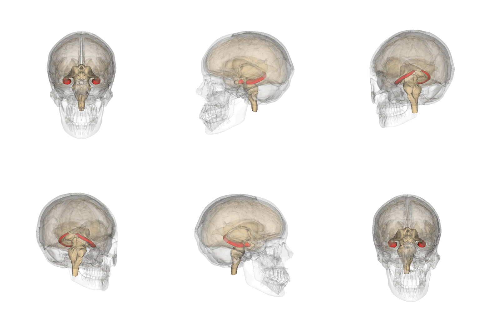
Hippocampus
There is a theory that claims that there is an intermediate stage between actual navigation and the maps from the hippocampus — the transformation of these maps into a first-person perspective. That is, a person tries to understand where things are not in general (as we see on real maps), but where things will be located relative to themselves (similar to the 'street view' function in Google Maps).
The authors of the studied work emphasize the following: Cognitive maps of the environment are encoded in the hippocampal formation in an allocentric system, while motor skills (the movements themselves) are represented in an egocentric system.
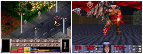
UFO: Enemy Unknown (allocentric system) and DOOM (egocentric system).
The difference between allocentric and egocentric systems is reminiscent of the difference between third-person games (or side-view, top-down, etc.) and first-person games. In the former case, the environment itself is important to us, while in the latter, our position relative to that environment matters. Thus, allocentric navigation plans must be converted into an egocentric system for actual implementation, i.e., movement in space.
Researchers believe that it is the dorsomedial striatum (DMS)* that plays a crucial role in the above process.
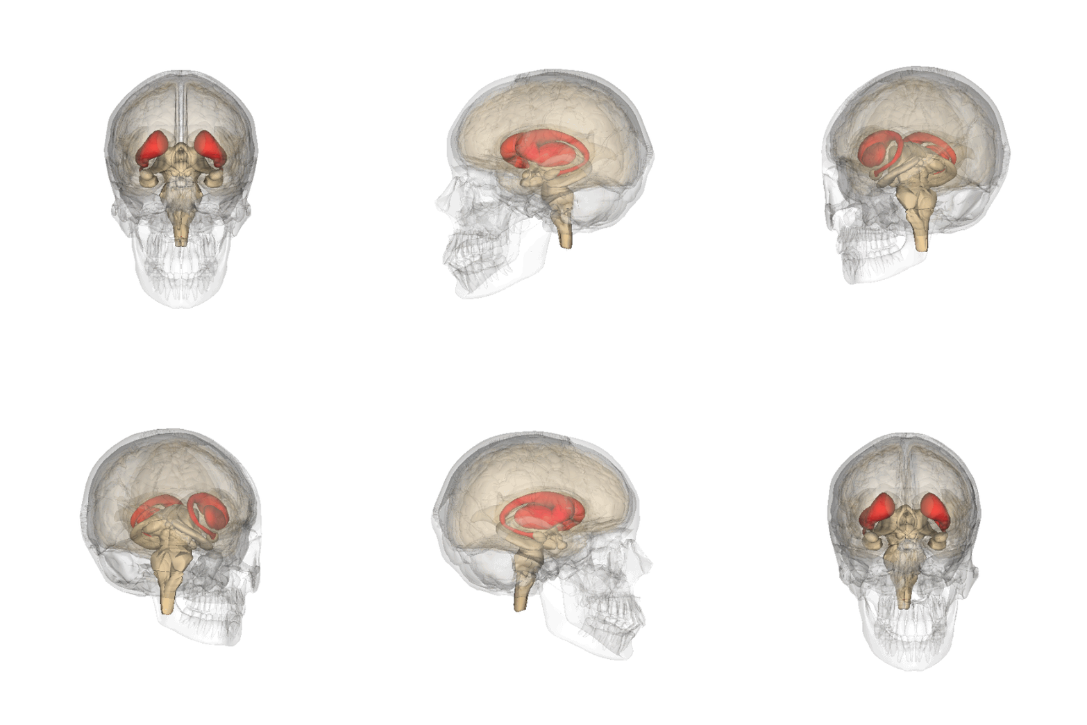
The striatum of the human brain.
Striatum* is a part of the brain that belongs to the basal ganglia; the striatum is involved in regulating muscle tone, internal organs, and behavioral responses; it is also referred to as the "striped body" due to its structure of alternating bands of gray and white matter.
The DMS demonstrates neuronal responses related to decision-making and actions regarding navigation in space, thus this area of the brain should be studied in more detail.
Research Results
To determine the presence/absence of egocentric spatial information in the striatum (DMS), 4 male rats were implanted with up to 16 tetrodes (special electrodes connected to specific areas of the brain), aimed at the DMS (1a).
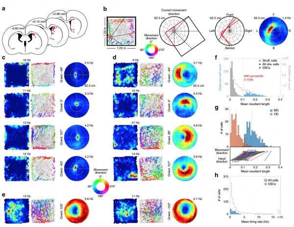
Image No. 1: response of cells in the striatum to boundaries of the environment in the egocentric reference system.
Explanation for Image No. 1:a — locations of the tetrodes;
b — egocentric map of boundaries;
with — allocentric spatial maps (4 squares on the left), trajectory graphs with color coding of peak cell reaction locations relative to body position and egocentric maps (4 squares on the right) based on the EBC cell reactions at different orientations and distances between the rat and the wall;
d — as well as on 1c, but for EBC with preferred distances away from the animal;
e — as well as on 1c, but for two reverse EBC;
f — distribution of the average resultant length for the observed cells;
g — distribution of the average resultant length for EBC using movement direction and head direction;
h — distribution of the average response of cells (all and EBC).
44 experiments were conducted where rats collected randomly scattered food in a familiar environment (open, not in a maze). A total of 939 cells were recorded. From the collected data, 31 head direction cells (HDC) were identified, but only a small portion of the cells, specifically 19, had allocentric spatial correlates. The activity of these cells, confined to the perimeter of the environment, was only observed when the rat moved along the walls of the test chamber, suggesting an egocentric scheme for encoding spatial boundaries.
To assess the capabilities of such an egocentric representation based on peak activity measures of the cells, egocentric maps of boundaries (1b), which illustrate the orientation and distance of the boundaries relative to the direction of the rat's movement, rather than the position of its head (comparison on 1g).
18% of the recorded cells (171 out of 939) exhibited significant responses when the boundary of the chamber occupied a certain position and orientation relative to the subject (1f). The scientists named them egocentric boundary cells (EBC — egocentric boundary cells). The number of such cells in the subjects ranged from 15 to 70, with an average of 42.75 (1c, 1d).
Among the egocentric boundary cells, there were those whose activity decreased in response to the boundaries of the chamber. There were a total of 49, and they were named reverse EBC (iEBC). The average response measure of the cells (their action potentials) in EBC and iEBC was quite low — 1.26 ± 0.09 Hz (1h).
The population of EBC cells responds to all variations of the orientation and position of the chamber boundary relative to the subject, but the distribution of preferred orientation is bimodal with peaks located at 180° opposite each other on either side of the animal (-68° and 112°), being slightly shifted from perpendicular to the long axis of the animal by 22° (2d).
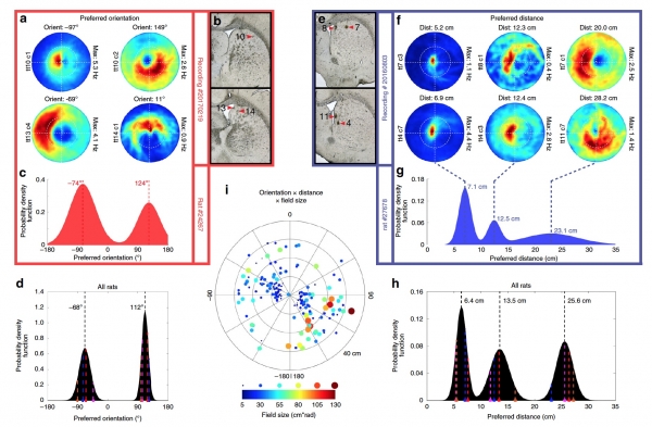
Image #2: preferred orientation and distance for the response of egocentric boundary cells (EBC).
Explanations for Image #2:a — egocentric boundary maps for four simultaneously studied EBCs with different preferred orientations indicated above each graph;
b — position of the tetrodes relative to the cells from 2a (numbers indicate the tetrode number);
with — distribution of probabilities of preferred orientations for all EBCs of one rat;
d — distribution of probabilities of preferred orientations for EBCs of all rats;
e — position of the tetrodes for the cells shown in 2f;
f — egocentric boundary maps for six simultaneously recorded EBCs with various preferred distances indicated above each graph;
g — distribution of probabilities of preferred distance for all EBCs of one rat;
h — distribution of probabilities of preferred distance for EBCs of all rats;
i — polar plot of preferred distance and preferred orientation for all EBCs, with space size represented by color and diameter of points.
The distribution of preferred distance to the boundary contained three peaks: 6.4, 13.5, and 25.6 cm, indicating the existence of three different preferred distances between EBCs (2f—2h), which may be important for a strategy of hierarchical navigational search. The size of the receptive fields of EBCs increased with the preferred distance (2i), indicating an increase in the accuracy of egocentric representation of the boundaries as the distance from the wall to the subject decreased.
Both in preferred orientation and distance, there was no clear topography, as active EBCs of the subject with various orientations and distances relative to the wall appeared on the same tetrode (2a, 2b, 2e and 2f).
It was also found that EBCs consistently respond to boundaries of space (walls of the chamber) in any variant of test chambers. To confirm that EBCs respond to local chamber boundaries rather than its distal features, the researchers "rotated" the position of the chamber by 45° and made several walls black, differentiating it from that used in previous tests.
Data was collected both in a normal testing chamber and in a rotated one. Despite the change in the testing chamber, all preferred orientations and distances relative to the walls for EBC subjects remained the same.
Given the significance of angles, the possibility that EBC uniquely encodes these local environmental attributes was also considered. By isolating the difference between responses near corners and those near the center of the wall, a subset of EBC cells (n = 16; 9.4%) was identified, which showed an enhanced response to corners.
Thus, it can be concluded that EBC cells respond significantly to the perimeter of the chamber, specifically to the walls of the testing chamber and its corners.
Next, the researchers checked whether the EBC cell response to open space (a testing arena without a maze, i.e., just 4 walls) was consistent across different sizing of the test area. Three trials were conducted, with each trial differing in wall length by 50 cm from the previous one.
Regardless of the size of the testing chamber, EBC responded to its boundaries at the same distance and orientation relative to the subject. This indicates a lack of scaling in response depending on the size of the environment.
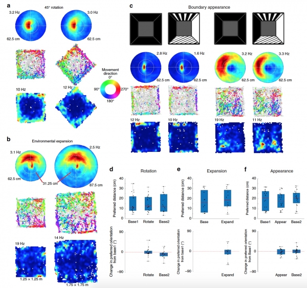
Figure 3: Stable response of EBC cells to spatial boundaries.
Explanations for Figure 3:a — Egocentric maps of EBC under normal conditions (left) and with the testing chamber rotated 45° (right);
b — Egocentric maps of EBC for a chamber size of 1.25 x 1.25 m (left) and for an enlarged chamber of 1.75 x 1.75 m (right);
with — Egocentric maps of EBC with normal black walls of the chamber (left) and with patterned walls (right);
d—f — Graphs of preferred distance (top) and changes in preferred orientation relative to the baseline (bottom).
Since the striped body receives information about the environment from multiple areas of the visual cortex, researchers also investigated whether the appearance of the walls (3c) of the chamber affected the response of EBC cells.
Changes in the appearance of spatial boundaries had no effect on the response of EBC cells or on the required distance and orientation for the response concerning the subject.
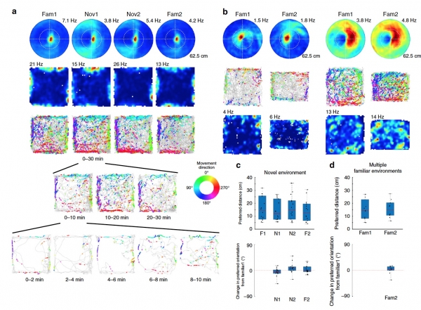
Figure 4: Stability of EBC cell response regardless of the environment.
Explanations for Image No. 4:a — egocentric maps for EBC in familiar (left) and new (right) environments;
b — egocentric maps for EBC obtained in the same environment but over a time interval;
with — graphs of preferred distance (top) and changes in preferred orientation relative to the baseline (bottom) for new (unfamiliar) environments;
d — graphs of preferred distance (top) and changes in preferred orientation relative to the baseline (bottom) for previously studied (familiar) environments.
It was also established that the response of EBC cells, as well as the necessary orientation and distance relative to the subject, do not change over time.
However, this "temporal" test was conducted in the same testing chamber. It was also necessary to check what the difference is between the EBC response to known conditions and new ones. For this purpose, several trials were conducted where rats explored a chamber they already knew from previous tests, followed by new chambers with open space.
As you may have guessed, the response of EBC cells + the required orientation/distance remained unchanged in the new chambers (4a, 4c).
Thus, the response of EBC provides a stable representation of the environmental boundaries relative to the subject in all types of this environment, regardless of the appearance of the walls, the size of the testing chamber, its movement, and the time spent by the subject in the chamber.
For a more detailed understanding of the nuances of the research, I recommend checking out the and related to it.
Epilogue
In this work, the researchers were able to practically confirm the theory of egocentric representation of the environment, which is crucial for spatial orientation. They demonstrated that there is an intermediate process between allocentric spatial representation and actual behavior, involving specific cells in the striped body, called egocentric boundary cells (EBC). It was also established that EBC are more related to the control of whole-body movement rather than just the head of the subjects.
This study aimed to identify the complete mechanism of spatial orientation, along with all its components and variables. According to the researchers, this work will further enhance navigation technologies for autonomous vehicles and robots, enabling them to understand the space around them as we do. The researchers are extremely pleased with the results of their work, which provide a foundation to continue exploring the relationship between specific areas of the brain and how navigation in space is performed.
Thank you for your attention, stay curious, and have a great working week, everyone! 🙂
Thank you for staying with us. Do you enjoy our articles? Would you like to see more interesting materials? Support us by placing an order or recommending us to your friends. 30% discount for Habr users on a unique entry-level server designed by us for you: (options available with RAID1 and RAID10, up to 24 cores and up to 40GB DDR4).
Dell R730xd for half the price? Only with us in the Netherlands! Dell R420 — 2x E5-2430 2.2GHz 6C 128GB DDR3 2x960GB SSD 1Gbps 100TB — from $99! Read about how
Source: habr.com
